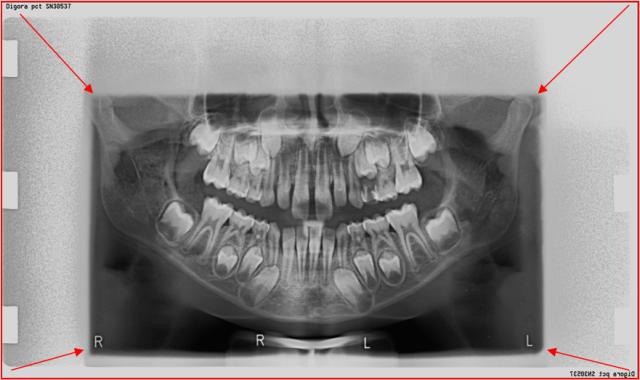
The recommended technique with a significantly reduced radiation field. The arrows indicate the reduction of the radiation field from the standard size of 15×30 cm and not by reducing the image. The upper parts of the facial bones and the cervical part of the spinal cord are not exposed to unnecessary radiation. Such an image can be digitally enlarged so that the blank area is not visible. By applying the digital method of the aforementioned technique, the exposure of children to X-rays is reduced by approximately 50-70%.
SPECIAL OPG (SCANOGRAMS of selected regions):
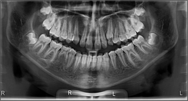
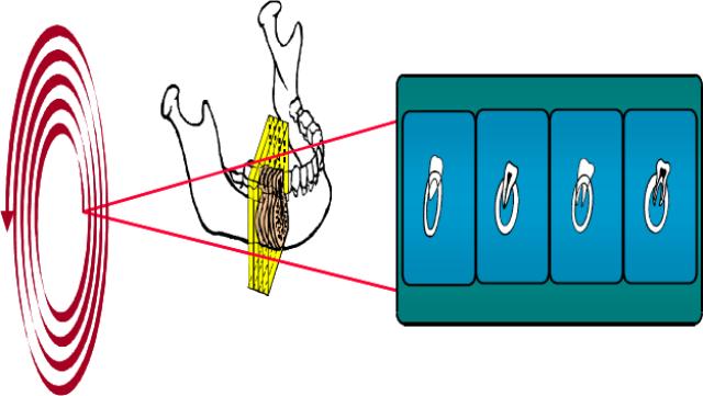
Scanographic imaging is essentially tomographic, only the orthopan tube is stationary at the moment of exposure
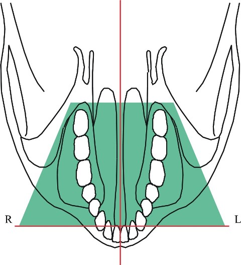
It is necessary to target the area in question • The alveolar extension of the upper and lower jaws • The maxillary sinus • The TM joint
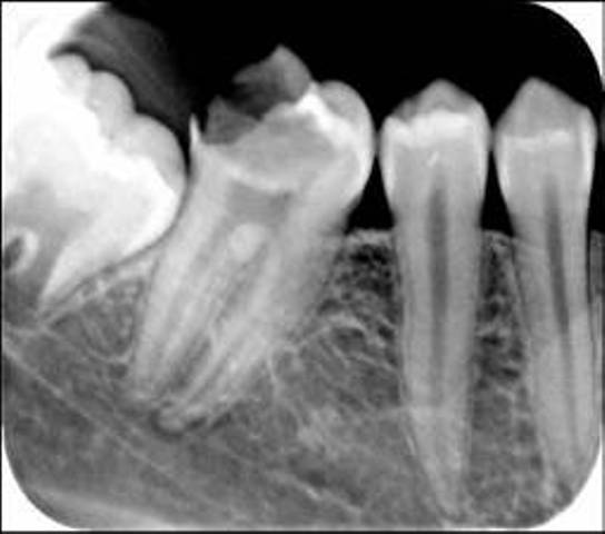
Since a very tight and strictly collimated beam of X-rays is focused, we get a sharper, less deformed image with higher contrast in the zone of dentition than with a large OPG. At the same time this technique of alveolar extensions replaces a large number of retroalveolar images.
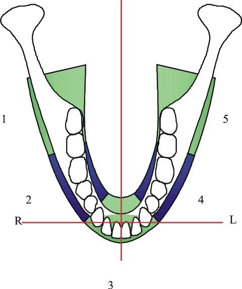
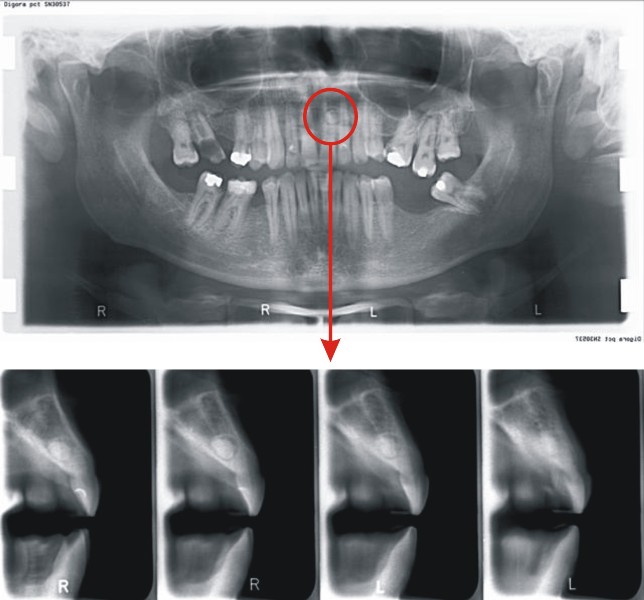
• Zone 1 – encompasses the right ramus and the upper and lower right molars • Zone 2 – upper-right molars and paramolars • Zone 3 – upper and lower front • Zone 4 – left upper and lower premolars and molars • Zone 5 – the left ramus and molars
A digital scanogram with zones 1-3-5 and 2-4
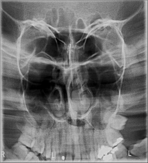
Maxillary sinuses
SCANOGRAM – of maxillary sinuses with clearly visible frontal sinuses, orbital, nasal septum, conchae, …
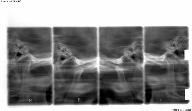
SCANOGRAM using the TMZ technique open – closed mouth
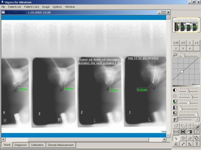
TOMOGRAPHY OF REGIONS SELECTED FOR IMPLANTOLOGY (cross - sectional)
(4 sagittal images in 2 or 4 mm layers)
The L – R labels are not valid
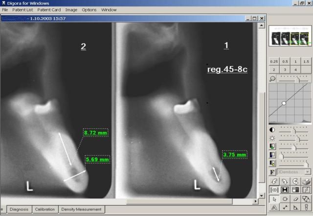
Cross sectional – tomography
Precise localization of the mesiodens in the sagittal section indicates a palatinal approach in the surgical intervention
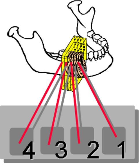
4 layers in 2 and 4 mm tomographic sections
The first on the right, image no. 1, is always the mesial layer, each subsequent layer is distal – numbers 2, 3 and 4
TELERADIOGRAPHS
RADIOGRAPHS OF FACIAL BONES
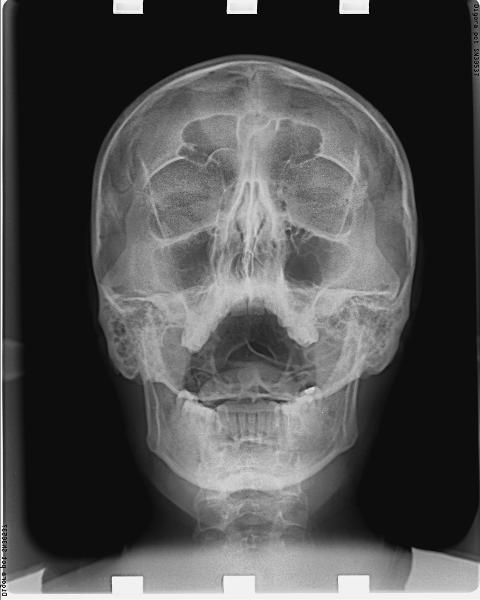
P – A Paranasal cavities
TELE RTG – lateral cephalogram
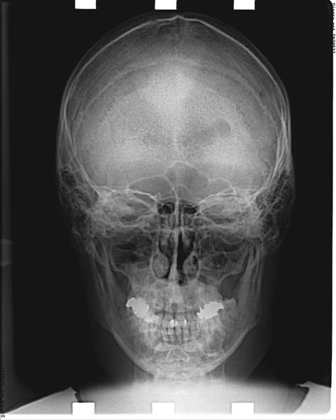
P – A Craniogram
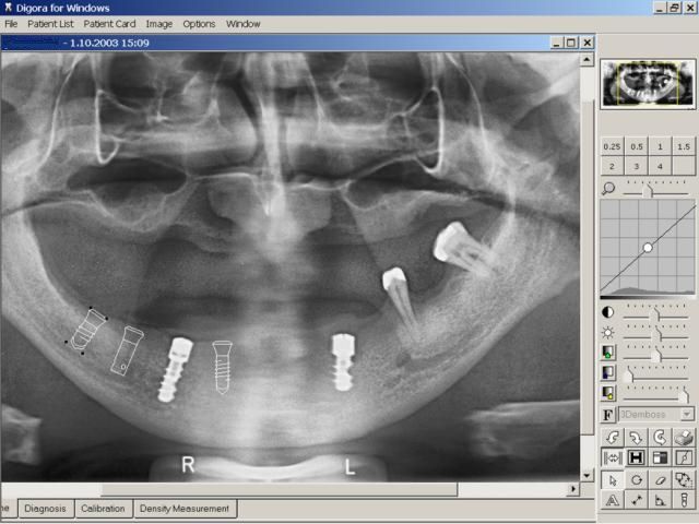
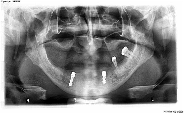
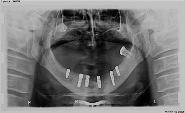
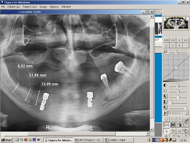
ANALYZING THE DIMENSIONS OF THE ALVEOLAR EXTENSION AND BONE DENSITY FOR THE PURPOSE OF IMPLANT PLANNING
So far, it has been impossible in dental radiography to achieve the kind of X-ray quality offered by spiral RTG tomography. It allows the therapist to analyze the images on a computer screen at a price up to 5 times lower than what the patient would have to pay for a CT image
Unsuccessful implanting on rtg. 33
After corrections – the restored state
In case of the analysis and measurement (of the mandibular corpus, for example) on an OPG image it is compulsory to take a tomographic cross section image which shows perfectly accurate information dimension-wise in the sagittal and vertical planes.
The selection of the region which will be tomographically imaged by the therapist can be designated on the referral by marking the region (e.g. 11, 12, …)
Tomography on a toothless maxilla
Tomography on the lower jaw in metric units
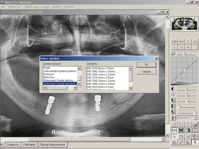
On the “Symbols” icon, it is possible to select various implant manufacturers – Straumann, Frialit, Branemark, etc. as well as all the possible dimensions of the implants with their original manufacturer’s codes.
The selected implant can be positioned in the desired region by dragging it with the mouse (drag and drop) along with the use of the “rotate” function.
DIGITAL TELE RTG ANALYSES:
Tele – RTG analyses with selected points, curves and angles, standardized or done according to the therapist’s selection. For a computer analysis of a digital image – an orthodontic craniometric analysis, it is sufficient to mark the craniometric points in order to obtain the desired analysis with a printout of the image, graphic and numeric values.
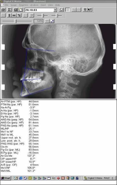
Burstone analysis
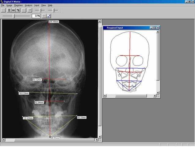
PA Cephalogram with an analysis in the transversal plane
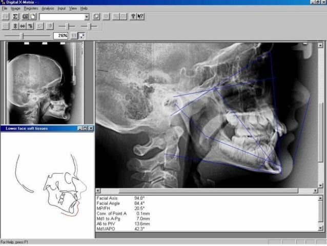
LAT Cephalogram with analyses of the vertical and saggital planes
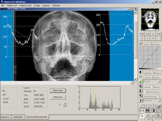
DENSITOMETRY:
The processing of bone density in a localized osteosclerotic change. It aids the diagnosis of osteoporosis, osteosclerosis, as well as is determining the growth type of long bones. The graph was determined in the marked zone, juxtaposed with a radiologically healthy zone for the purpose of comparison.
ALL THE AFOREMENTIONED IMAGING TECHNIQUES CAN BE ANALOG OR DIGITAL AND PROCESSED USING ORIGINAL SOFTWARE
AND MAY AT YOUR REQUEST BE SENT TO YOUR COMPUTER VIA THE INTERNET.
THE IMAGES ARE ON A REAL METRIC SCALE WITH A SUB mm PRECISION
The CANAL of the mend. nerve in reg. 35 – sagittal at 2 mm, measured details of the distance to the mandib. canal, the width of the mandib. body, the width of the canal lumen, etc.
PRECISE SUB-mm MEASUREMENT
Carried out after calibrating, that is re-scaling to a real scale, proportionate to real dimensions because each technique has a certain magnification factor.
DIGITAL IMAGES ARE PRINTED OUT ON PHOTOGRAPHIC PAPER AND RECORDED ON CDs
TELEDENTISTRY:
With the development of information technology and telecommunication it has become possible to create an internal network between therapists of different specialties, whereby it would be much faster to exchange results, referrals, X-rays, details on the course of the treatment and all other information for the purpose of additional consultations on possible therapies. This allows a continuity in further specialization which takes place on a daily basis in the course of dental practice.